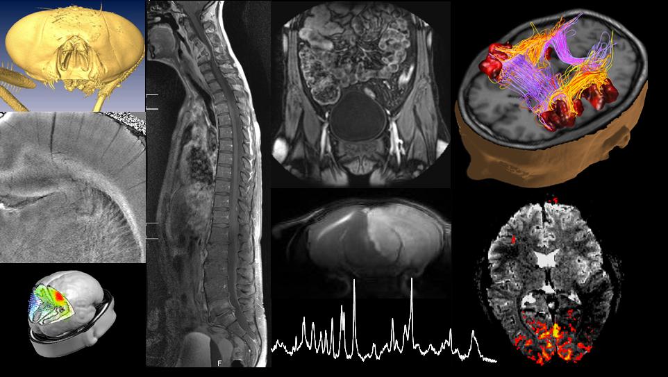
Fundamentals of biomedical imaging
Weekly outline
-
Welcome !
This course covers the physical principles of major in vivo bio-imaging modalities, i.e., ultrasound, computed tomography, emission computed tomography, positron emission tomography and magnetic resonance imaging. We will show how existing physical principles transcend into bio-imaging and establish an important link into life sciences, illustrating the contributions physics can make to life sciences.
In the lectures, practical examples will be shown to illustrate the respective imaging modality, its use, premise and limitations, and biological safety will be touched upon.
The student will develop a good understanding of the mechanisms leading to tissue contrast of the bio-imaging modalities covered in this course, including the inner workings of the scanner and how they define the range of possible biomedical applications.
Based on this course, the student will be able to judge which imaging modality is adequate for specific life science needs and to understand the limits and promises of each modality.
The course was live broadcast in 2014 (and partially in 2015), using google+ hangout on air, the videos are available also on our dedicated youtube channel, FundBioImag. The links to the recorded courses are provided on this site in the content description of the respective week.
-
Indications on what is required for the exam
Location: PO 01, 22.06.2019 time 12:15-15:15
-
1: Introduction (pdf)
- Course organization
- Bio-Imaging in Life sciences
- Essential elements of bio-imaging (Webb Ch. 5.3 & 5.4)*
- Lab visit
*indicates approximate correspondence with the book of Andrew Webb, but course may be more extensive or more qualitative. It is not recommended to use the book other than as additional support.
Following this week you
- know the course organization and coverage of topics;
- know the contribution of bio-imaging to life science and why it is an interdisciplinary effort.
- know the main elements required for bio imaging;
- are able to perform contrast to noise and signal to noise calculations;
- are familiar with noise error propagation calculations
-
The quizzes are here for training and are complementary to exercises; the "marks" during quizzes are only indicative and do not count for the final grade!
- Course organization
-
2: Ultrasound and Introduction to x-rays (pdf)
- Introduction to ultrasound imaging (Ch. 3.8, 3.10)
- Ionizing radiation
- Generation of ionizing radiation (Ch. 1.2)
Following this week you
- understand the basic principle of ultrasound imaging
- Are able to estimate the influence of frequency on resolution and penetration
- are capable of calculating echo amplitudes based on acoustic impedance;
- know which parts of the electromagnetic spectrum are used in bio-imaging
- know the definition of ionizing radiation;
- understand the principle of generation of ionizing radiation and control of energy and intensity of x-ray production;
-
The quizzes are here for training and are complementary to exercises; the "marks" during quizzes are only indicative and do not count for the final grade!
- Introduction to ultrasound imaging (Ch. 3.8, 3.10)
-
3: Interaction of x-rays with tissue (pdf)
- Linear attenuation coefficient (1.4)
- Interaction of photons with matter (1.5)
Rayleigh scattering
Compton Scattering
Photoelectric effect
Pair production- Effective atomic number Zeff (1.7)
- Radioprotection - a short primer (1.14)
Following this week you
- know the definition of linear and mass attenuation coefficient;
- understand the major mechanisms of x-ray absorption in tissue;
- understand the dependence of these mechanisms on photon energy and tissue composition;
- are able to perform contrast-to-noise calculations using effective Z;
- are able to apply the basic principles of radiation protection.
-
The quizzes are here for training and are complementary to exercises; the "marks" during quizzes are only indicative and do not count for the final grade!
-
4: From x-ray to image: Computed tomography (pdf)
- Experimental principle (1.10)
Beam hardening effects
Sensitivity and resolution considerations- Intensity projection (1.11)
Radon Transform
Projection reconstruction (App. B)
Sinogram- Central slice theorem
Following this week you
- understand the cause of beam hardening and its influence on image contrast;
- are familiar with the Radon transform, the pre-requisite for x-ray image reconstruction;
- understand the principle of matrix reconstruction and backprojection;
- are able to perform simple matrix image reconstruction tasks
- understand the limits and potential of CT.
-
The quizzes are here for training and are complementary to exercises; the "marks" during quizzes are only indicative and do not count for the final grade!
-
5: Emission computed tomography (pdf)
- Experimental principle (2.1)
Tracers
- Resolution issues: collimators (2.7)
- Attenuation correction (2.9)
- x-ray detection - scintillation followed by amplification (2.7)
- Scatter correction (2.9)
Following this week you
- understand the reason for collimation in imaging g-emitting tracers and its implication on resolution/sensitivity;
- understand the implications of x-ray absorption on emission tomography;
- are familiar with the basic principle of radiation measurement using scintillation;
- are familiar with the principle/limitations of photomultiplier tube amplification;
- understand the use of energy discrimination for scatter correction.
-
The quizzes are here for training and are complementary to exercises; the "marks" during quizzes are only indicative and do not count for the final grade!
-
6: Positron Emission Tomography (2.11) (pdf)
- Coincidence detection - electronic collimation
- Scatter and random correction
- Attenuation correction
- Factors affecting resolution
After this course you
- are capable of describing the essential elements of a PET scan
- can distinguish the principle of PET detection from that of SPECT
- understand the bases of scatter elimination and attenuation correction
- understand the factors affecting spatial resolution in PET.
- understand the reason why PET/CT has become the industry standard.
-
The quizzes are here for training and are complementary to exercises; the "marks" during quizzes are only indicative and do not count for the final grade!
-
7: Tracer kinetics (pdf)
- Compartment models
- Fick's principle
- One-tissue compartment model
- Modeling tracer uptake
- deoxyglucose uptate
Following this week you
- understand how mass conservation can be used to estimate metabolic rates
- understand the mathematical principle underlying metabolic modeling of imaging data
- can apply the principles of modeling tracer uptake to simple kinetic situations
- understand the basics of modeling deoxyglucose uptake into tissue to extract metabolic rates
-
The quizzes are here for training and are complementary to exercises; the "marks" during quizzes are only indicative and do not count for the final grade!
-
8: Introduction to magnetic resonance (4.1, 4.2, 4.4) (pdf)
- The MR scanner
- Magnetism of the nucleus (spin)
- The basic equation of motion of MR: precession
- Rotating frame of reference
After this course you
- are familiar with the prerequisites for nuclear spin
- know the factors determining nuclear magnetization
- Can compare magnetization for different nuclei and magnetic fields
- Know the equation of motion for magnetization
- Are able to describe the motion of magnetization in lab and rotating frame
- Understand that MRI has complex mechanisms and contrasts
-
The quizzes are here for training and are complementary to exercises; the "marks" during quizzes are only indicative and do not count for the final grade!
-
Happy Easter !

-
Enjoy the Easter Break !

-
9: Relaxation of nuclear magnetization (4.2) (pdf)
- Detection (and excitation) of the MR signal
- Rotating frame revisited
- Resonance: effective RF field and flip angle
- Relaxation
- T1 and T2
- Bloch equations
- Relaxation basics: free induction decay (FID)
After this week you
- Can describe principle of MR detection and excitation
- Can explain how MR excitation is frequency selective (resonance)
- Understand the principle of relaxation to the equilibrium magnetization
- Know what are the major relaxation times and how they phenomenologically affect magnetization in biological tissue, in particular that of water.
- Can explain the elements of the Bloch equations and FID
- Understand that MR contrast strongly depends on experimental parameters
-
The quizzes are here for training and are complementary to exercises; the "marks" during quizzes are only indicative and do not count for the final grade!
-
10: NMR spectroscopy (4.10) (pdf)
Practice session today in BS 160
After this week you
1. can calculate the effect of multiple RF pulses on longitudinal magnetization
2. know the definition of Ernst angle
3. Understand the two basic mechanisms by which electrons influence the precession frequency of nuclear magnetization
4. Know the definition of chemical shift
5. Know how and under what molecular conditions NMR spectroscopy can provide non-invasive biochemical information-
The quizzes are here for training and are complementary to exercises; the "marks" during quizzes are only indicative and do not count for the final grade!
-
11: Echo formation and spatial encoding (4.3) (pdf)
- The 3rd magnetic field: Gradient
- Spatial encoding
- Gradient dephasing and rephasing
Gradient echo sequence
k-spaceFollowing this week you
- Understand the principle of slice selection
- Are familiar with dephasing and rephasing of transverse magnetization and how it leads to echo formation
- Understand the principle of spatial encoding in MRI
- Can describe the basic imaging sequence and the three necessary elements
- Understand the principle of image formation in MRI and how it impacts spatial resolution
-
The quizzes are here for training and are complementary to exercises; the "marks" during quizzes are only indicative and do not count for the final grade!
-
12: MRI contrast mechanisms (pdf)
- T2*-weighted contrast: BOLD fMRI (4.11)
- Hahn spin echo (4.2)
- T1-, T2- and proton-density weighted MRI
- MRI contrast agents (4.7)
After this weeks course you
- are capable of describing the biophysical basis of BOLD contrast
- Understand the mechanism of spin echo generation
- Know the three contrasts that can be generated by the spin echo imaging sequence and how the timing parameters are optimized for each contrast
- Understand why the same tissue appear bright on T2 weighted images and dark on T1 weighted images
- Understand the mechanism by which the two principal contrast agent mechanisms lead to signal increase or decrease
.-
The quizzes are here for training and are complementary to exercises; the "marks" during quizzes are only indicative and do not count for the final grade!
-
13: Magnetic resonance: Advanced contrast mechanisms & Overview of imaging modalities (pdf)
- Effect of flow
- Diffusion-weighted MRI (4.9)
- Anisotropic diffusion
Limitations
- Comparison by examples
Après ce cours vous
- Understand the influence of motion on the phase of magnetization
- Understand how random motion leads to echo amplitude reduction
- Are able to calculate the attenuation of the MR signal due to diffusion
- Understand how diffusion-weighted MRI signal reflects cellular structure and how this can be exploited to track nerve fibers, among others
- Have a firm grasp on the premises and limitations of the imaging modalities covered in this course
-
The quizzes are here for training and are complementary to exercises; the "marks" during quizzes are only indicative and do not count for the final grade!